Virus: Definition, structure and functions


This post includes the definition and structure of virus in detail. Now, first of all, we define virus……………….. “The virus is a submicroscopic extremely complex infectious agent, that is nonliving molecules, that can grow and reproduce only in living cells” Typically a layer of protein around a core of genetic material of RNA or DNA is present in the virus structure. It does not have any semipermeable membranes, and cause several important diseases in humans, animals, and plants.
They are most notorious because of their pathogenic nature. When we discuss viruses, Ebola outbreak in West Africa (2014), and the 2009 H1N1/swine flu pandemic likely comes in our mind. More recently coronavirus respiratory disease pandemic (COVID-19) in 2020 has caused a great threat all over the world economy. But viruses are research tools also, for further understanding of the basic cellular processes such as the mechanics of protein synthesis, and of viruses themselves.


Virus structure
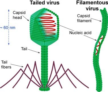
The following three basic components are included in the virus structure. The first two are present in all types of viruses. But the last one viral envelope is present mainly in the animal virus structure.
- A nucleic acid genome
- A protein capsid that covers the genome. (Genome plus protein capsid is called the nucleocapsid).
- Lipid envelop (In many animal viruses)
All the intact virus is called a virion. Detail of these components is right below.
A- Viral genomes:
Although the genomes of all known cells are made up of double-stranded DNA, the genomes of viruses can be made up of single-stranded or double-stranded DNA or RNA. Viruses vary greatly in their size, ranging from approximately 5-10 kb (Papovaviridae, Parvoviridae, etc.) to even more than 100-200 kb (Herpesviridae, Poxviridae). Different structures of virus genomes are given below.
1- DNA
- Double-stranded – linear or circular
- Single strand – linear or circular
- Other structures – gapped circles
2- RNA: double-stranded – linear
Single-stranded linear structure: These single-chain genomes can be positive-sense single-stranded RNA or negative-sense single-stranded RNA or ambisense. The positive-sense single-stranded RNA can directly serve as mRNA and encode proteins, so for these viruses, viral RNA is infectious. The negative-sense single-stranded RNA is not infectious, as it must be copied into the + sense strand before it can be translated. In an Ambisense virus, some part of genome is sense strand and some part is the antisense.
The genome of some RNA viruses is segmented, which means that a virus particle contains several different RNA molecules, like different chromosomes.
B: Protein capsid
Protein capsid is an essential component of virus structure. Viral genomes are surrounded by shells of proteins known as capsids. An interesting question is how the capsid proteins recognize viral RNA or DNA, but not cellular DNA. The answer is that there is often some kind of “packaging” signal (sequence) in the viral genome that the capsid proteins recognize. A capsid is composed of repetitive structural subunits that are arranged in one of two symmetrical structures, a helix or an icosahedron. These “subunits” may consist of a single polypeptide, in simplest cases. However, in many cases, these structural subunits (also called protomers) are made up of various polypeptides. The detail of helical and icosahedral virus structures is described below.
1) Helical capsids: The first and best example studied is the plant tobacco mosaic virus (TMV), which contains a single-stranded RNA genome and a protein coat made up of a single 17.5 kd protein. This protein is arranged in a helix around the viral RNA, with 3 nucleotides of RNA fitting into a groove in each subunit. The helical capsids can also be more complex as they involve more than one protein subunit.
A Helix can be defined by two parameters, its diameter, and pitch. This structure is very stable and can be easily disassociated and re-associated by changing the ionic strength, pH, temperature, etc. The interactions that hold these molecules together are not covalent and involve H bonds, salt bridges, hydrophobic interactions, and Vander Waals forces.
Several families of animal viruses contain helical nucleocapsids, for example, Orthomyxoviridae (influenza), Rhabdoviridae (rabies) and Paramyxoviridae (bovine respiratory syncytial virus). These are all enveloped viruses.
2) Icosahedral capsids: In these structures, the subunits are arranged in the form of a hollow, quasi-spherical structure, with the genome inside. An icosahedron is defined as consisting of 20 equilateral triangular faces arranged around the surface of a sphere. They exhibit 2-3-5 symmetry.
- 2-fold rotational symmetry through the edges.
- 3-fold rotational symmetry through the center of each triangular face.
- 5-fold symmetry through the center of each corner.
These corners are also called vertices, and each icosahedron has 12.
Since proteins are not equilateral triangles, each side of an icosahedron contains more than one protein subunit. The simplest icosahedron is made using 3 identical subunits to form each face, so the minimum number of subunits is 60 (20 x 3). Remember that each of these subunits could be a single protein or, more likely, a complex of multiple polypeptides.
Many viruses have a genome too large to be packaged within an icosahedron made up of only 60 polypeptides (or even 60 subunits), making them even more complicated. But in these, the total number of subunits is always a multiple of 60.
Apparent clusters or “Lumps” are often seen on the surface of the particle, when viral nucleocapsids are viewed under the electron microscope, These are generally protein subunits grouped around an axis of symmetry and have been called “morphological units” or capsomeres”
C: Viral Envelope
In some animal viruses, the nucleocapsid is surrounded by a membrane, also called an envelope. This envelope is made up of a lipid bilayer and is made up of lipids from host cells. It also contains virus-encoded proteins, often glycoproteins that are transmembrane proteins. These viral proteins serve many purposes, such as binding to receptors in the host cell, playing a role in membrane fusion and cell entry, etc. They can also form channels on the viral membrane.
Many enveloped viruses also contain matrix proteins, which are internal proteins that bind the nucleocapsid to the envelope. They are very abundant (that is, many copies per virion) and are generally not glycosylated. Some virions also contain other non-structural proteins that are used in the viral life cycle. Examples of this are replicases, transcription factors, etc. These nonstructural proteins are present in low amounts in the virion.
Enveloped viruses are formed by budding through cell membranes, usually the plasma membrane, but sometimes an internal membrane such as ER, Golgi, or nucleus. In these cases, the assembly of viral components (genome, capsid, matrix) occurs on the inner side of the membrane, the envelope glycoproteins clump together in that region of the membrane, and the virus breaks out. This ability to bud allows the virus to leave the host cell without lysing or killing the host. Conversely, unenveloped viruses and some enveloped viruses kill the host cell to escape.
Characteristics of viruses
Living characters:
- They reproduce at a fantastic speed, but only in living host cells. e.g. in bacterias, humans, animals, and plants “
- They can mutate their genetic material and so are able adaptable in changing environments.
Non-living characters:
- We can define virus as the acellular particles which are without cytoplasm, cell membrane, nucleus organelles, etc.
- They do not metabolize, grow or divide, on their own and require metabolic machinery of the host cell to replicate. Instead, the new viral components are assembled within the infected host cell.
- Virus structure includes one or two strands of DNA or RNA but not both.
- Viruses are covered with a protective protein layer called as CAPSID.
- They are inactive when they are not inside a living cell, but they are active when they are inside another living cell.
Interesting Facts about viruses
- Viruses are not classified in any of the five kingdoms of living things. This means that they are not bacteria, fungi, protists, plants or animals.
- Most viruses are so small that they cannot be seen under a light microscope.
- The word “virus” comes from the Latin word “virulentus” which means“a poisoned wound” or “full of poison.”
- Viruses can sometimes attack and kill bacteria (Bacteriophages).
- The first human virus discovered was the yellow fever virus in 1901 by Walter Reed.
- A virus that contains RNA instead of DNA is sometimes called a retrovirus.
- There are two main types of reproductive cycles for viruses: the lytic cycle and the lysogenic cycle.
- Diseases caused by a virus with a lytic cycle show symptoms much faster than viruses with a lysogenic cycle.
Related posts:
Recent Posts
Types of Cells
Living things are made up of cells, the basic unit of life. There are many types…
What is the Difference Between HIV and AIDS?
The difference between HIV and AIDS is that AIDS is the disease caused by HIV infection. You…
What is Difference between Antisepsis and asepsis?
The Difference between Antisepsis and asepsis is that The antisepsis is the procedure performed to reduce…
Virus vs bacteria
Virus vs bacteria: The difference between viruses and bacteria lies in the fact that the virus…
What is Difference between Arches and bacteria?
Major Difference between Arches and bacteria is that The archaea and bacteria are prokaryotes, unicellular living whose genetic material…
What is The Difference Between DNA and RNA?
The difference between DNA and RNA is that DNA is deoxyribonucleic acid and RNA is…
